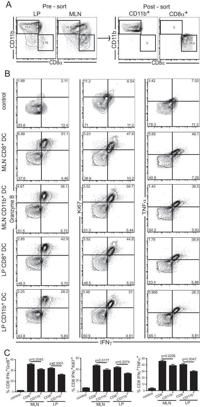
FIG 5.
In vitro stimulation of immune CD8 T cells by mucosal DC subsets. CD11b+ CD8− or CD11b− CD8+ DC subsets were isolated at day 2 p.i. from the MLN of infected C57BL/6 mice or the LP of infected C57BL/6 mice treated with FTY720. (A) Cell suspensions from the LP and MLN before (left) and after (right) sorting are shown after gating on live CD11c+ cells. (B) Magnetically enriched CD8+ T lymphocytes from infected IFN-γ great mice were stimulated overnight with CD11b+ CD8− or CD11b− CD8+ DC from either the LP or the MLN. Dot plots showing IFN-γ, granzyme B (GrzB), TNF-α, and Ki67 expression are gated on live CD8αβ cells. The negative control corresponds to nonstimulated BMDC (no antigen) incubated with immune CD8+ T cells. (C) Graphs represent the frequencies of double-positive CD8+ T cells (IFN-γ+ GrzB+, IFN-γ+ Ki67+, and IFN-γ+ TNF-α+) for the corresponding subsets. Data represent results of at least 2 experiments with 3 mice/group.

