FIG 6.
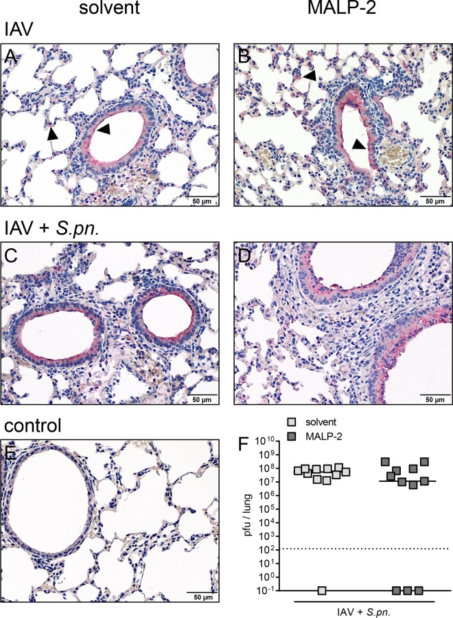
The pulmonary IAV load remained unaltered by MALP-2 stimulation. Mice infected with 102 PFU IAV were treated with 0.5 μg MALP-2 or solvent on day 5 and challenged with 103 CFU S. pneumoniae or PBS on day 6. Seven days after IAV infection, lung sections were prepared, and immunohistochemistry for IAV (red staining) was performed. IAV was observed mainly within bronchi and in alveolar macrophages (black arrowheads) of lungs from both solvent (A and C)- and MALP-2 (B and D)-treated mice. (E) Lung sections from sham-infected and solvent-treated mice served as a negative control. Representative images are shown (n = 3 or 4). (F) Lung viral loads after secondary bacterial infection were determined on day 7. Values are given as individual data and means (n = 11). The dotted line indicates the lower detection limit.

