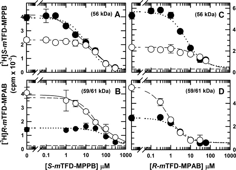
FIGURE 10.
S-mTFD-MPPB and R-mTFD-MPAB inhibition of S-[3H]mTFD-MPPB or R-[3H]mTFD-MPAB photolabeling of α1β3γ2 GABAARs. GABAARs were photolabeled by S-[3H]mTFD-MPPB (A and C) or R-[3H]mTFD-MPAB (B and D) in the presence of bicuculline (●, 30 μm) or GABA (○, 300 μm) and nonradioactive S-mTFD-MPPB (A and B) or R-mTFD-MPAB (C and D). 3H incorporation into the 56-kDa (A and C) or 59/61-kDa (B and D) subunit bands was determined by liquid scintillation counting. Data are average (± S.D.) of two separate experiments. Data were fit to single-site (nH = 1, dot or dash traces) or two-site (solid traces) binding models as described under “Experimental Procedures.” Parameter fits are tabulated in Table 2.

