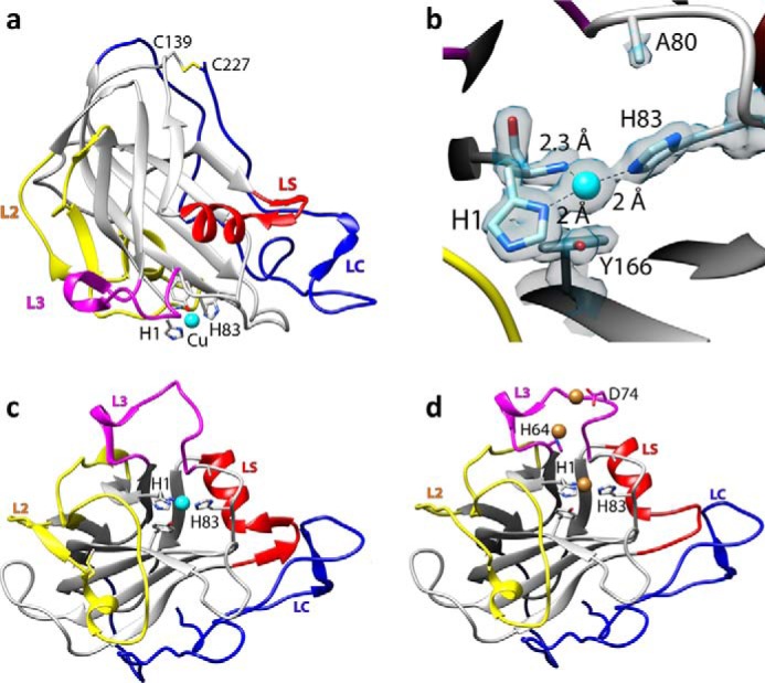
FIGURE 1.

Structural representations of NcLPMO9C-N. a, cartoon representation of the copper-bound structure; copper is depicted as a cyan sphere; b, close up of the copper-binding site with the electron density map around the active site in gray mesh (contoured at 1σ); c, overall structure of the copper-loaded protein rotated by 90° along the horizontal axis compared with the view in a; d, structure of the zinc-loaded protein with the three bound zinc ions depicted as brown spheres; the orientation is similar to that in c. Note the structural variation in the loop coordinating the third zinc ion (residues 70–76 in the L3 loop, colored in pink).
