FIGURE 4.
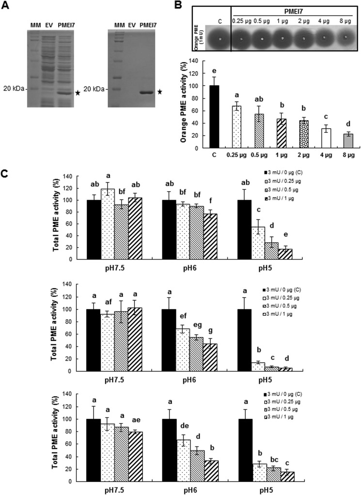
Expression of PMEI7-His6 in bacteria. Purification and tests of PME activity inhibition. A, expression of PMEI7-His6 in E. coli and purification. SDS-PAGE analysis of total protein extracts (left) and Ni-NTA-purified protein extracts (right) of isopropylthio-β-galactoside-induced cultures containing empty vector (EV) or recombined vector (PMEI7). MM, molecular mass markers. B, gel diffusion assay of the inhibitory capacity of the purified PMEI7-His6 on commercial orange PME at pH 6.0. Experiments were carried out using 1 milliunit of orange PME and various quantities of purified PMEI7-His6. Results are means ± S.D. (error bars) of six replicates. The different letters indicate data sets significantly different according to Tukey's range test, preceded by a one-way ANOVA having p < 0.001. C, quantification of the pH dependence of the inhibitory capacity of PMEI7-His6 on total PME activity of cell wall-enriched protein extracts from three Arabidopsis organs. Top, 3-week-old light-grown leaves; middle, 10-day-old light-grown roots; bottom, 4-day-old dark-grown hypocotyls. 3 milliunits of total PME activity was used with either PMEI7-His6 storage solution (■) or a PME activity/PMEI7-His6 (μg) ratio of 3:0.25 (dotted bars), 3:0.5 ( ), and 3:1 (▨). Results are means ± S.D. of six replicates. The different letters indicate data sets significantly different according to Tukey's range test, preceded by a one-way ANOVA having p < 0.001.

