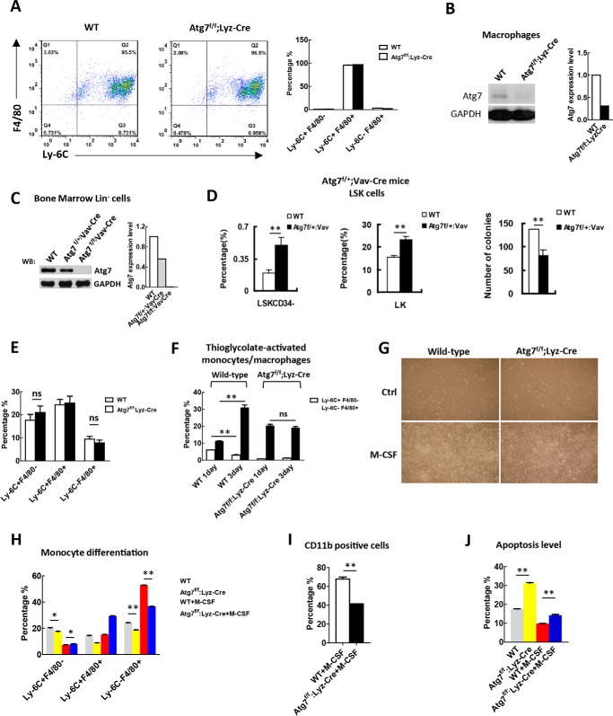
FIGURE 2.
Atg7 deletion in myeloid cells cripples the artificial but not physiological induction of monocyte-macrophage differentiation. A, sorting for myeloid cells at the monocyte-macrophage differentiation stage (Ly6C+F4/80+). Bone marrow cells from Atg7f/f;Lyz-Cre mice and wild-type mice were sorted against the markers with FACS, shown by representative sorting plots (left). The purity of the sorted cells from these two mouse models is shown (right). B, examination of the efficiency for atg7 gene floxing in macrophages of the mouse models as indicated, and the majority of atg7 protein is absent due to the floxing of atg7 gene. To obtain sufficient macrophages for analysis, macrophages were sorted with FACS after M-CSF induction of mononuclear cells isolated with MACS from bone marrow cells. Shown is a representative Western blotting result (left) and quantified data (right). C, examination of the efficiency for atg7 gene floxing in HSPCs of Atg7f/f;Vav-Cre mice and Atg7f/+;Vav-Cre mice by Western analysis (left), and their protein levels were quantified (right). Monoallelic deletion of atg7 only partially reduced atg7 protein level but biallelic deletion caused complete absence of atg7 protein in HSPCs. D, monoallelic deletion of the atg7 gene in Atg7f/+;Vav-Cre mice caused hematopoietic stem cell exhaustion. Percentage of LSKCD34− and LK cells were measured by flow cytometry (left and middle panels). Colony-forming ability of LSK cells from wild-type mice and Atg7f/+;Vav-Cre mice were determined, and the result was quantified (right panel). E, flow cytometric analysis of the percentage of monocytes (Ly-6C+F4/80−) and macrophages (Ly-6C−F4/80+) from bone marrow cells of Atg7f/f;Lyz-Cre mice and wild-type mice. F, examination of artificial induced monocyte-macrophage differentiation by thioglycolate in Atg7f/f;Lyz-Cre mice and wild-type mice. Differentiation was measured by flow cytometry on days 1 and 3 after induction. G, morphological changes associated with or without M-CSF (25 ng/ml)-induced monocyte-macrophage differentiation. H–J, analysis of differentiation and apoptosis of the atg7-deleted monocytes. Primary monocytes from Atg7f/f;Lyz-Cre and wild type mice were sorted against the indicated markers and incubated with or without M-CSF (25 ng/ml) for 72 h, the differentiation and apoptosis levels of monocytes were measured by flow cytometry. Data are representatives or statistical results of three experiments. n ≥ 6. *, p < 0.05; **, p < 0.01.

