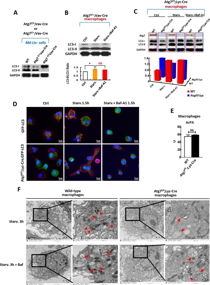
FIGURE 3.
Macrophages but not hematopoietic stem and progenitor cells maintain autophagic response when atg7 is deleted. A, effect of monoallelic or biallelic deletion of atg7 gene in stem and progenitor cells on autophagy response. Western analysis on HSPCs from homogenous Atg7f/f;Vav-Cre mice, Atg7f/+;Vav-Cre heterozygous mice and wild-type mice show that biallelic deletion of agt7 gene in HSPCs caused complete loss of LC3-II conversion from LC3-I, but monoallelic deletion of atg7 gene maintained the LC3 lipidation and processing. B, effect of monoallelic deletion of atg7 gene directed by Vav promoter in macrophages on autophagy response. Western analysis on macrophages from heterozygous Atg7f/+;Vav-Cre mice shows that monoallelic deletion of agt7 gene caused loss of canonical autophagic flux response, albeit constitutive LC3-II conversion from LC3-I maintained. For starvation, serum was deprived for 90 min. For inhibition on atg7-dependent canonical autophagy, bafilomycin A1 of 10 nm was used. The upper panel is a representative Western blotting result, and the lower panel is quantified results from three independent experiments. C, effect of biallelic deletion of atg7 gene directed by Lyz promoter in macrophages on autophagy response. The primary macrophages were lysed after being plated in the culture plate for 2 h under starvation or with/without Baf-A1 (10 nm) treatment and were then immunoblotted with antibodies against Atg7 and LC3 (upper panel). Autophagic flux was quantified (lower panel). Shown is a representative result of three independent experiments. GAPDH served as a loading control in experiments A, B, and C. D, confocal microscopic analysis of GFP-LC3 localization in the macrophages of Atg7f/f;Lyz-Cre;GFP-LC3 mice and GFP-LC3 transgenic mice. The GFP-LC3 transgenic mice served as a control. Representative images are shown for non-starvation (left panel) and for starvation 1.5 h (middle panel) with or without 10 nm of Baf-A1 (right panel). The nucleus was stained with DAPI (blue), CD11b (red) is a marker for macrophages. GFP-LC3 (green) is expressed in all tissue cells including macrophages of the GFP-LC3 transgenic mice. E, macrophages acid phosphatase activity (AcPA) in wild-type and Atg7f/f;Lyz-Cre mice was measured by spectrophotometry. F, transmission electron microscopic analysis of macrophage autophagy in response to starvation treated with or without bafilomycin A1 (Baf, 10 nm), a specific inhibitor on atg7-dependent canonical autophagy. Macrophages were isolated from wild-type mice or Atg7f/f;Lyz-Cre mice and followed by starvation for 3 h. Autophagosomes and autolysosomes are indicated with arrows. Data are representatives or statistical results of three experiments. n≥6. *, p < 0.05; **, p < 0.01.

