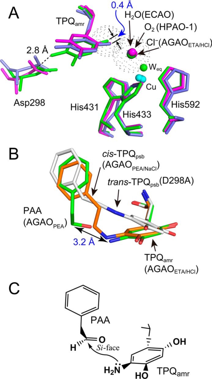
FIGURE 11.

Effect of binding of chloride ion at the axial position of the copper center on the conformation of TPQamr. A, conformation of the TPQamr ring in the AGAOETA/HCl structure (green) is compared with those of the substrate-reduced E. coli CAO (ECAO) (purple) and HPAO-1 (magenta). Cyan spheres, copper atoms. Residue numbers are referred to those of AGAO. van der Waals surfaces of the chloride ion and the oxygen atom of the 2-OH group of TPQamr are represented with gray dots. B, comparison of cis-TPQpsb formed in AGAOPEA/NaCl (orange), cis-TPQpsb formed in the D298A mutant of AGAO (29) (gray), TPQamr formed in AGAOETA/HCl (green), and PAA formed in AGAOPEA (green). C, schematic drawing of the presumed mechanism of the formation of cis-TPQpsb in AGAOPEA/NaCl.
