FIGURE 4.
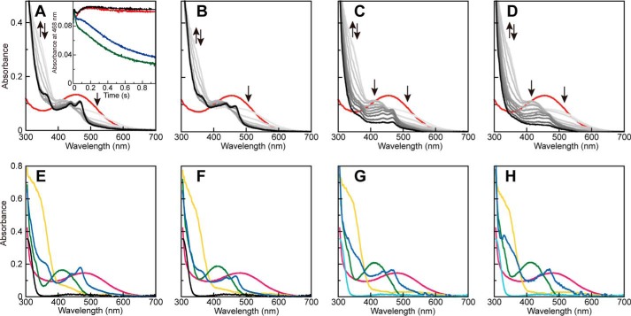
Spectral changes during the reductive half-reaction with 2-PEA. UV-visible absorption spectra were recorded after mixing AGAO (100 μm monomer) with 2 mm 2-PEA (A), 2 mm 2-PEA plus 300 mm Na2SO4 (B), 2 mm 2-PEA plus 100 mm NaCl (C), or 2 mm 2-PEA plus 300 mm NaBr (D) in 50 mm HEPES buffer, pH 6.8, at 4 °C under anaerobic conditions. The spectra obtained at 0 (red), 2.30, 3.84, 6.4, 8.96, 14.1, 24.3, 44.8, 117, 209, 332, 600, and 1023 ms are shown using darker colors to represent later times. The arrows indicate the direction of the spectral changes. Deduced absorption spectra are shown for TPQox (red), TPQssb (yellow), TPQpsb (green), TPQamr (black), and TPQsq (blue) in E, F, G, and H, calculated from the measurements of A (no salt), B (300 mm Na2SO4), C (100 mm NaCl), and D (300 mm NaBr), respectively. The absorption spectra for TPQamr·X− (cyan) generated during the experiments using 100 mm NaCl (C), and 300 mm NaBr (D) are shown in G and H, although they are defined to be identical to that of TPQamr (black). The inset of A shows the time course of the absorbance at 468 nm in the spectral changes of A (no salt, red), B (300 mm Na2SO4, black), C (100 mm NaCl, blue), and D (300 mm NaBr, green).

