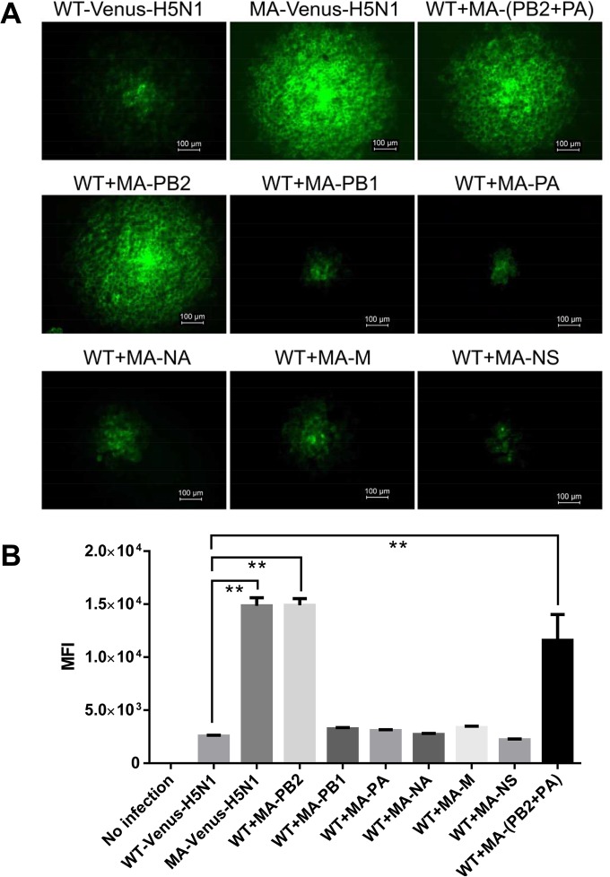
FIG 2.
Venus expression of various H5N1 viruses in MDCK cells. (A) MDCK cells were infected with Venus-H5N1-related viruses, and at 24 hpi, the Venus expression of each virus plaque was observed by using fluorescence microscopy (Axio Observer.Z1; Zeiss). A representative image of each virus is shown. (B) MDCK cells were infected with Venus-H5N1-related viruses at an MOI of 0.001, and at 24 hpi, MDCK cells were digested with trypsin into single cells and analyzed by flow cytometry. The mean fluorescence intensity (MFI) was calculated by using FlowJo X 10.0.7r2. The values are means ± standard deviations from two independent experiments. **, P < 0.01 compared with WT-Venus-H5N1 virus-infected cells.

