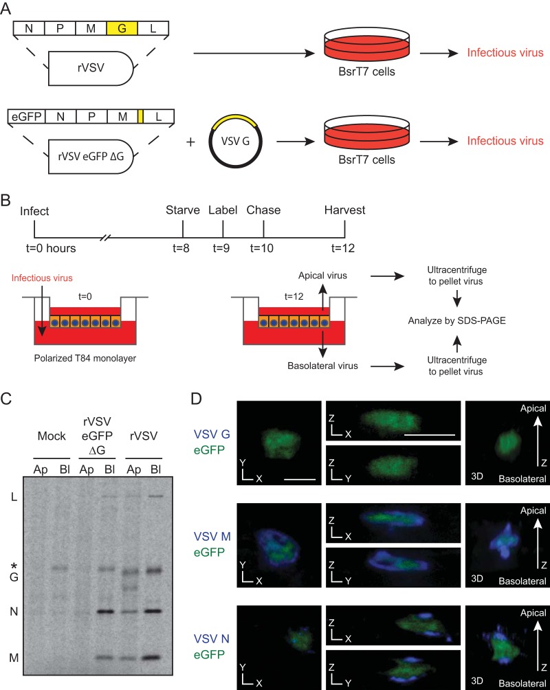
FIG 2.
Test of the requirement of G for directional budding. (A) Schematic for growth of rVSV and rVSV eGFP ΔG with G supplied in trans. (B) Time line of infection, radiolabeling, and collection. (C) Concentrated virus from 12 hpi was analyzed by SDS-PAGE. Note that 1/20 of the rVSV, relative to rVSV eGFP ΔG, from basolateral (Bl) budding was loaded for clarity. The basolateral lane of rVSV eGFP ΔG contains a cellular band (*) with mobility similar to that of G that also appears after mock infection. Both rVSV and rVSV eGFP ΔG had significantly more N in the basolateral chamber (Student's t test P values of <0.01 and <0.05, respectively; n = 5). Ap, apical. (D) T84 cells were infected with rVSV eGFP ΔG and analyzed by confocal microscopy for the proteins indicated. Cross sections and three-dimensional renderings are shown. The lack of G was verified by immunocytochemistry analysis, and consequent defective virus production was confirmed by titration (data not shown). The scale bars represent 10 μm.

