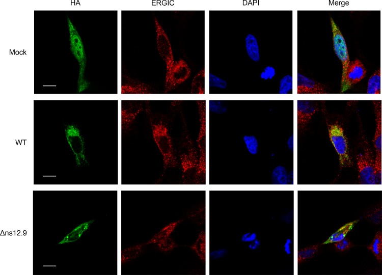
FIG 7.
Subcellular localization of ns12.9 during HCoV-OC43 infection. RD cells were infected with HCoV-OC43-WT or HCoV-OC43-Δns12.9 at an MOI of 1. At 24 h postinfection, cells were transfected with pCAGGS-ns12.9-HA. Cells were fixed and examined by confocal microscopy analysis at 24 h posttransfection. ns12.9 was stained with anti-HA monoclonal antibody and visualized with Alexa Fluor 488-conjugated goat anti-mouse antibody (green). ERGIC was stained with ERGIC53 antibody (H-245) and visualized with Cy3-conjugated goat anti-rabbit antibody (red). Nuclei were stained with DAPI (blue). Bars represent 20 μm.

