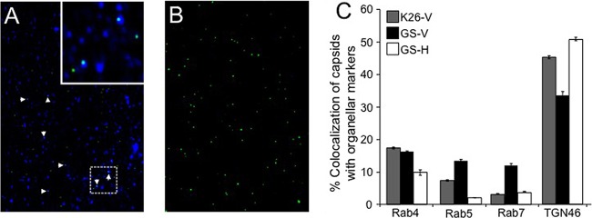
FIG 2.
HSV capsids colocalize with organellar markers in the absence of UL36p. Membrane-associated fractions were isolated from K26GFP or HSV1-GS2491-infected Vero or HS30 cells. They were then attached to coverslips, immunostained with anti-TGN46, anti-Rab4, anti-Rab5, or anti-Rab7 antibodies and appropriate secondary antibodies, and visualized in the green (capsid) and Cy5 (antibody) channels. (A) Representative field of HSV1-GS2491 (UL36-null) capsid-associated particles stained with anti-TGN46 antibodies. White arrowheads indicate colocalization of capsids (green) and TGN46 immunoreactivity (blue). The inset at the top right corner is a 3-fold magnification of the boxed region in the lower part of the panel. (B) Field similar to that in panel A but omitting anti-TGN46 antibodies. (C) Determination of percent colocalization based on the total number of VP26-GFP-positive K26GFP or HSV1-GS2491 particles compared to the number staining with each antiorganellar antibody (indicated below the graph). Viral strain and cell type are indicated in the key at the top left, labeled as in Fig. 1. The numbers of individual particles counted (n) for each sample were as follows: K26-V, n = 1,326; GS-V, n = 517; GS-H, n = 1,518. Plotted values represent means and standard deviations from the means.

