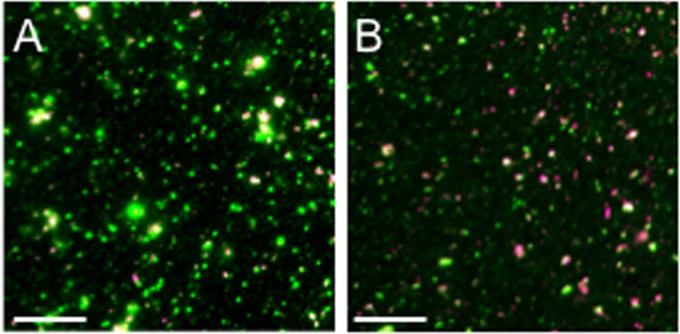
FIG 3.
Representative fields of fluorescent dye-stained cytoplasmic HSV particles. PNS was prepared from HSV1-GS2491-infected Vero (A) or HS30 (B) cells, and then virions and organelles were attached to a microscope slide and stained with the fluorescent lipophilic dye DilC18(5)-DS. After fixation, fields of particles were imaged in the green (VP26-GFP) and Cy5 (infrared, dye) channels; merged images are shown. GFP-positive/dye-positive and GFP-positive/dye-negative particles were counted to generate the data in Tables 1 and 2. Bar, 10 μm.

