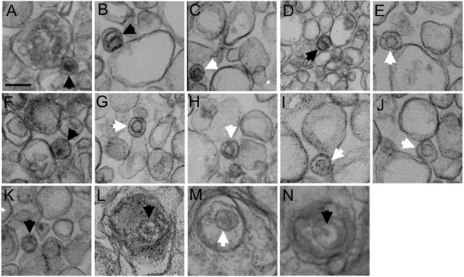
FIG 5.
Transmission electron microscopy of membrane-associated HSV particles. Vero (A to K) or HS30 cells (L to N) were infected with UL36-null HSV1-GS2491, and then cytoplasmic buoyant virus-associated organelles were prepared as described for Fig. 1C. The organelle-containing fraction was pelleted, fixed, and processed for thin-section electron microscopy. White and black arrowheads in each panel indicate B and C capsids, respectively. All images are at the same magnification. Bar in panel A, 100 nm.

