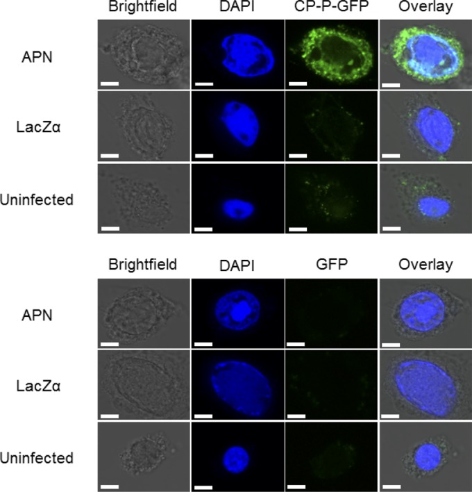
FIG 4.

Aminopeptidase N is necessary for internalization of CP-P-GFP. Visualization of CP-P-GFP endocytosed into APN-expressing Sf9 cells using confocal microscopy. Sf9 cells were infected with vAPN-GPI(+) or vLacZα. At 48 h postinfection, the cells were incubated with CP-P-GFP or GFP, followed by DAPI to stain the nuclei. Uninfected Sf9 cells served as an additional control. DAPI and intracellular GFP fluorescence were detected by the examination of multiple cell layers using a confocal microscope. Increased fluorescence was observed in Sf9 cells expressing recombinant APN. No fluorescence was observed when cells were incubated with GFP. The images are representative of two replicate experiments, with fluorescence seen in approximately half of the cells in the CP-P-GFP treatment. Scale bars, 5 μm.
