FIG 6.
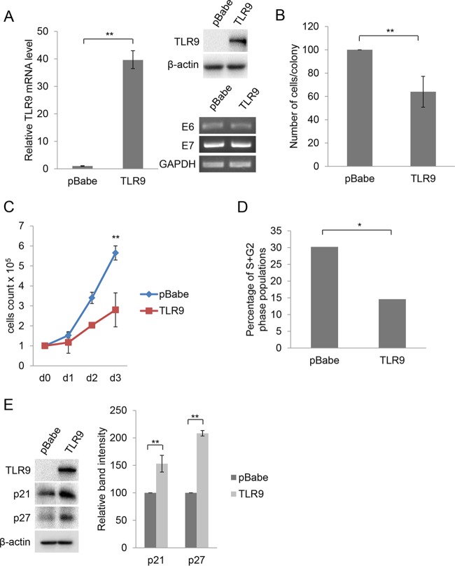
TLR9 expression in HPV38 E6/E7 HFK inhibits cellular proliferation by the accumulation of p21WAF1 and p27Kip1. (A) HPV38 E6/E7 HFK were transduced with pBabe empty vector or expressing TLR9. The efficiency of transduction was verified by RT-PCR (left) and IB (right). Data on the left are the means from three independent experiments. **, P < 0.01. The bottom right shows the levels of HPV38 E6 and E7 mRNA determined by qPCR. (B) Colony formation assay. After 6 to 8 days of culture in puromycin-containing medium, colonies were fixed in 20% methanol and stained with crystal violet. The number of cells per colony was determined by cell counting. Results are the mean counts from 10 colonies randomly selected from three independent experiments. Double-blind counting was performed. **, P < 0.01. (C) Growth time course. The growth of all cell populations was monitored for 3 days as described in Materials and Methods. The growth was assessed by counting live cells with trypan blue. Data represent the means from two independent experiments, each performed in duplicate. **, P < 0.01. (D) Flow cytometry analysis of cells described for panel C (day 3) stained by propidium iodide. Data are from one representative experiment of three independent experiments. *, P < 0.05. (E) Total proteins (20 μg) from HPV38 E6/E7 HFK transduced with pBabe or pBabe-TLR9 were analyzed by IB (left) and band intensities quantified and normalized to β-actin levels (right). Data are the means from three independent experiments. **, P < 0.01.

