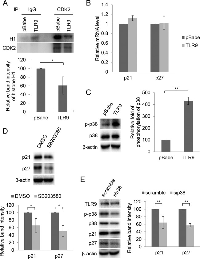
FIG 7.
TLR9-induced accumulation of p21WAF1 and p27Kip1 is mediated by activation of p38 signaling. (A) Kinase assay. CDK2 was immunoprecipitated from 1 mg of total proteins in whole-cell lysate. As a control, an identical reaction was carried out with a negative IgG antibody. Immunocomplexes were then incubated with purified histone H1 in the presence of [γ-32P]ATP. After SDS-PAGE followed by autoradiography (top), band intensity was quantified (bottom). Data are the means from three independent experiments. *, P < 0.05. (B) Total RNA from indicated cell lines was extracted and p21WAF1 and p27Kip1 expression levels were measured by RT-qPCR. Error bars represent standard deviations from four biological replicates. (C) Total proteins (20 μg) from HPV38 E6/E7 HFK transduced with pBabe or pBabe-TLR9 were analyzed by IB (left). The intensity of the protein bands in three independent experiments was quantified (right). **, P < 0.01. (D) TLR9-expressing HPV38 E6/E7 HFK were treated with the p38 inhibitor SB203580 (5 μM) or dimethyl sulfoxide (DMSO). After 3 h, total proteins were extracted and analyzed by IB using the indicated antibodies (top). The intensity of the protein bands in three independent experiments was quantified (bottom). *, P < 0.05. (E) TLR9-expressing HPV38 E6/E7 HFK were transfected with siRNA scramble or siRNA specific for p38 (sip38). After 48 h, total proteins were extracted and analyzed by IB (left). Band intensity was quantified in three independent experiments and is shown as a histogram (right). **, P < 0.01.

