
Figure 5.
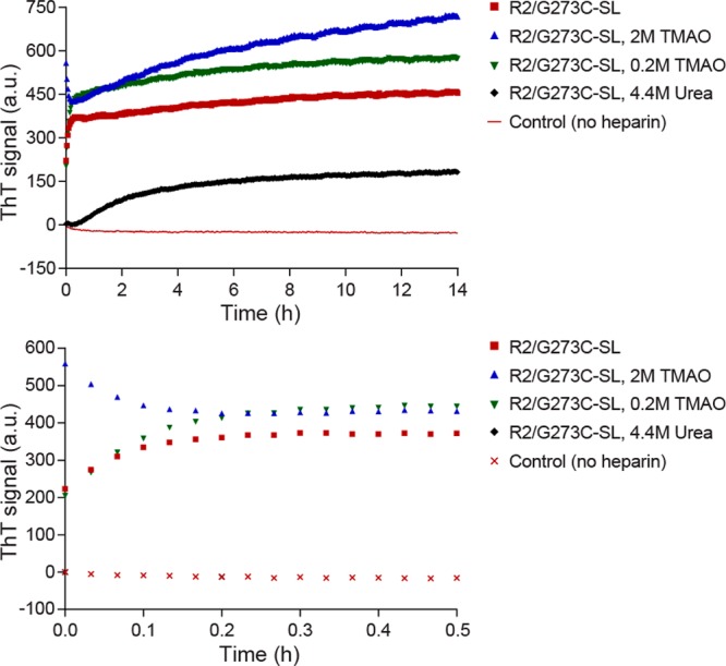
Thioflavin T fluorescence reveals effects of heparin and osmolytes on tau aggregation. Top panel is full scale while the bottom panel is zoomed in to show the first 30 min. The spin-labeled tau peptide R2/G273C-SL (25 μM) was incubated in 20 mM ammonium acetate buffer, pH 7.0, in the presence or absence of 6 kDa heparin (6.25 μM), TMAO (2 M), TMAO (0.2 M), and urea (4.4 M). All four non-heparin conditions generate identical and overlapping ThT signals, and so only one is shown for simplicity. Data shown are averaged from five separate experiments.