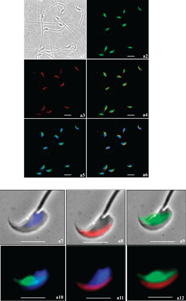
FIG. 6.
Immunofluorescence analysis of SPESP1 (green signal) in fixed, permeabilized, noncapacitated (a) and capacitated, acrosome-reacted (b) spermatozoa. Noncapacitated spermatozoa and acrosome-reacted spermatozoa both retained SPESP1 immunofluorescence, which was confined to the equatorial segment. Status and position of the anterior acrosome was imaged using PNA lectin (red signal). a) Noncapacitated sperm: a1) bright field; a2) SPESP1 immunofluorescence confined to equatorial segment; a3) PNA staining of anterior acrosome; a4) merged image of PNA-positive anterior acrosome and SPESP1-positive equatorial segment demonstrating distinct domains; a5) green SPESP1domain overlain by blue 4′,6-diamidino-2-phenylindole (DAPI) nuclear staining; a6) merged image of SPESP1, PNA, and DAPI domains; a7) dual bright-field/fluorescence images of blue DAPI-

