
FIG. 7.
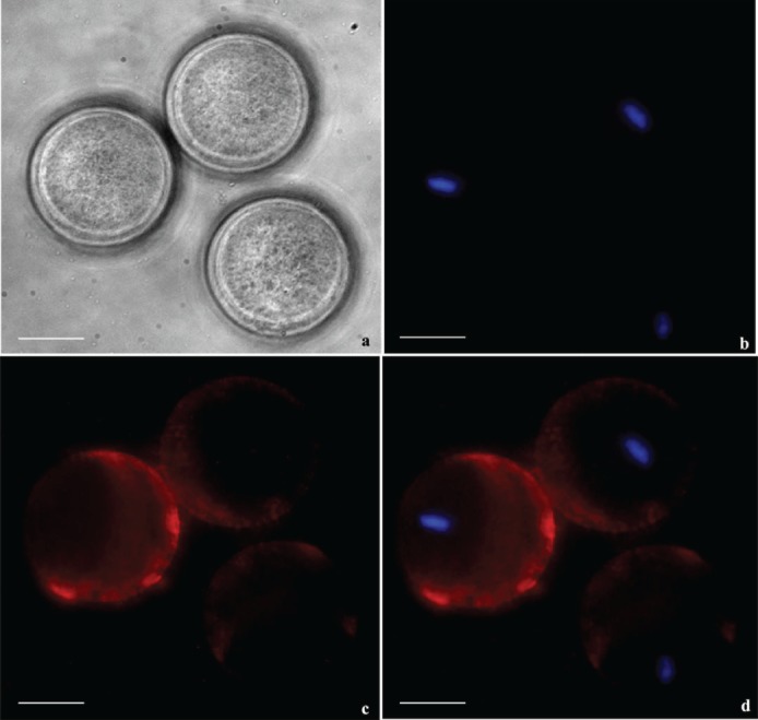
Zona-free mouse oocytes were incubated with recSPESP1 protein and probed with SPESP1 immune and nonimmune antibody to detect the oolemmal binding domain of SPESP1. Bright field (a), nuclear 4′,6-diamidino-2-phenylindole (DAPI) stain in blue (b), immune SPESP1 in red (c), and overlapped image (d) revealed SPESP1 binding to microvillar domain antipodal to eccentrically located M2 nucleus. Bar = 25 μm.