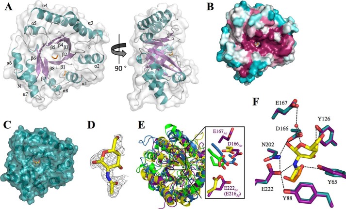
FIGURE 4.
Structure of Sph3Ac(54–304) and investigation of conserved regions. A, Sph3Ac shown in schematic representation with transparent surface model reveals a (β/α)8 barrel fold. The surface β-strands (purple) and α-helices (blue) are labeled β1–8 and α1–8, respectively. Loop regions are shown in light gray, and the ethylene glycol molecules are depicted as orange sticks. B, representation of the conserved surface as calculated by the ConSurf server (49–51) reveals a conserved groove on the top-face (C termini of β-strands) of the (β/α)8 barrel with a small depression in the center. Conserved residues are shown in fuchsia and variable residues in teal. C, transparent surface representation of the Sph3Ac structure (teal) in complex with GalNAc (yellow). D, close-up of the |Fo − Fo| omit density map contoured around GalNAc at σ = 2.5. E, superposition of the (β/α)8 barrels of Sph3 (purple) and a representative member of GH18 (blue, PDB 4WIW), GH27 (yellow, PDB 1KTB), and GH84 (green, PDB 2CBJ) shows the similarity in the overall folds. Right panel, close-up of the active sites of these glycoside hydrolases reveals three acidic residues of Sph3 (Asp-166, Glu-167, and Glu-222) in the same vicinity. E, comparison of the ethylene glycol molecule (orange) found in the active site of the apo-structure (purple) with GalNAc (yellow) from the co-crystal structure (teal). Residues connected with black lines represent those within hydrogen bonding distance.

