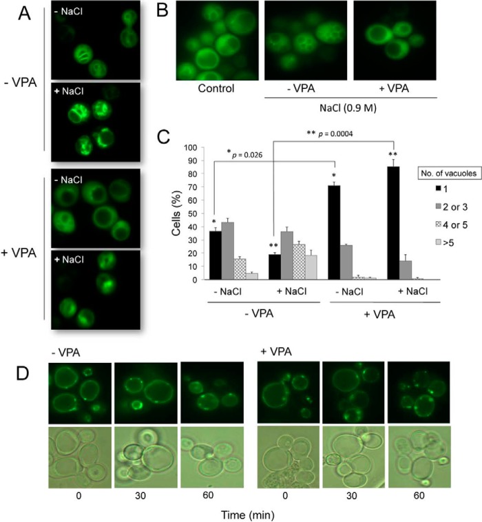
FIGURE 6.
PI3,5P2 is perturbed in VPA-treated cells. A, intensity of fluorescence associated with the PI3,5P2-binding probe is higher in untreated cells (−VPA) following exposure to osmotic shock (+NaCl). Wild-type cells constitutively expressing the GFP-tagged PI3,5P2-binding probe (Atg18-GFP) were grown in SM at 30 °C to the mid-log phase and treated with 2 mm VPA for 1 h. Cells were harvested and exposed to hyperosmotic stress for 5 min in SM to which NaCl was added to a final concentration of 0.9 m. Cells were observed by fluorescence microscopy. B, vacuolar fragmentation in response to hyperosmotic stress. C, vacuolar morphology of VPA-treated and -untreated cells was analyzed by fluorescence microscopy and quantified. The number of cells containing 1, 2–3, 4–5, or >5 vacuoles was determined. For each condition, about 100 cells were analyzed. The results are the mean of three independent experiments ± S.E. (error bars). Statistical analysis using a two-tailed unpaired t test indicated a significant effect of VPA on the number of cells with single vacuoles. *, p = 0.026 for (−NaCl), and **, p = 0.0004 for (+NaCl). D, Wild-type cells constitutively expressing a GFP-tagged PI3P-binding probe (FYVE-GFP) were grown in SM at 30 °C to the mid-log phase. VPA was added to a final concentration of 2 mm. Cells were harvested at the indicated times following addition of the drug and visualized by fluorescence microscopy.

