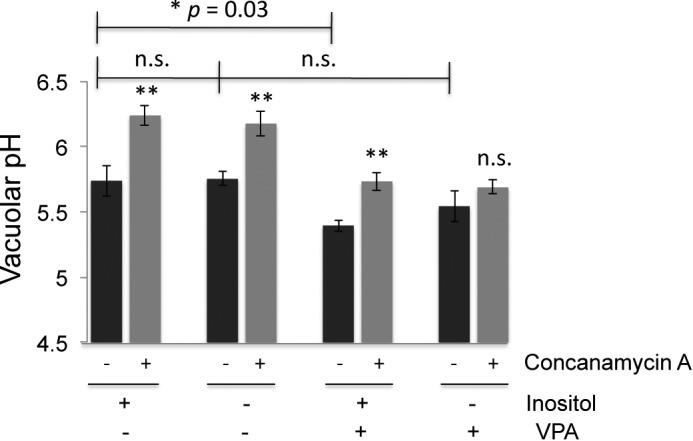
FIGURE 9.

Vacuolar pH was measured using BCECF. Cells were grown to log phase overnight in the indicated medium buffered to pH 5 with 50 mm potassium phosphate and 50 mm potassium succinate. Cells were briefly deprived of glucose, and then vacuolar pH was averaged over a period of 100 s, starting 8 min after glucose re-addition. Black bars represent the mean of four independent experiments, with error bars corresponding to the standard error of the mean. Gray bars correspond to the means of paired samples incubated with the V-ATPase inhibitor concanamycin A for 15 min before glucose re-addition. Statistical comparison (unpaired t test) of I+ samples grown overnight with and without VPA indicated a significant difference with p = 0.03. There was no significant (n.s.) difference between I+ and I− samples (p = 0.94) or I− samples grown with or without valproate present (p = 0.29). The average vacuolar pH in the presence and absence of concanamycin A was also compared for each sample. All of the samples except the I−, VPA+ sample showed highly significant differences between concanamycin-treated (gray bars) and -untreated (black bars) (**, p = 0.005–0.009). There was not a significant difference (p = 0.12) with concanamycin A treatment of the cells grown in I− medium with VPA.
