FIGURE 3.
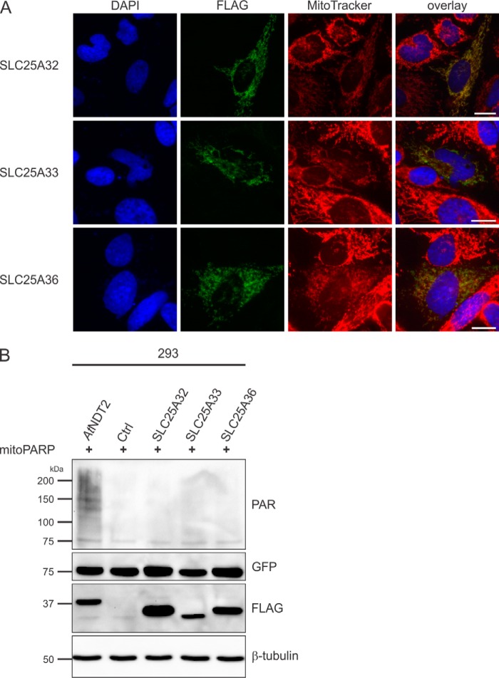
Human SLC25A32, SLC25A33, and SLC25A36 proteins do not exhibit mitochondrial NAD+ transporter activity. A, HeLa S3 cells were transiently transfected with plasmids encoding C-terminally FLAG-tagged human SLC25A32, SLC25A33, and SLC25A36 proteins. The images show the nuclei (DAPI) and mitochondrial structures (MitoTracker) and the overexpressed proteins (FLAG). As revealed by the merged images, the recombinant proteins co-localize with mitochondria. Scale bar, 10 μm. B, 293 cells were transiently co-transfected with a vector encoding mitoPARP and either control vectors or vectors encoding the indicated transporter. Cell lysates were subjected to PAR immunoblot analyses. The presence of AtNDT2 strongly increases polymer (PAR) formation indicative of elevated mitochondrial NAD+. The expression of the transporters (FLAG) and mitoPARP (EGFP) was confirmed, and β-tubulin served as a loading control. Ctrl, control.

