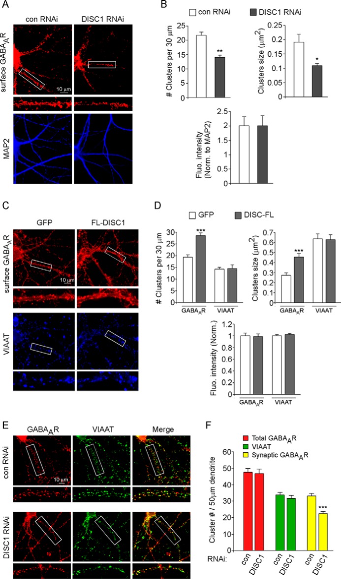
FIGURE 3.

DISC1 knockdown reduces surface and synaptic GABAAR clusters, whereas DISC1 overexpression increases surface GABAAR clusters. A and C, immunocytochemical images of surface GABAAR β2/3 subunits in cortical cultures transfected with a control (con) versus DISC1 shRNA (A) or GFP versus FL-DISC1 (C). Following surface GABAAR β2/3 labeling, neurons were permeabilized and co-stained with MAP2 (A) or VIAAT (C). Enlarged versions of the boxed regions of dendrites are also shown. B and D, quantitative analysis of surface GABAAR β2/3 or VIAAT clusters in cortical cultures with different transfections. ***, p < 0.001; **, p < 0.01; *, p < 0.05; Student's t test. Fluo, fluorescence; Norm, normalized. E, immunocytochemical images of control shRNA- versus DISC1 shRNA-transfected cortical neurons co-stained with GABAAR β2/3 and VIAAT. Enlarged versions of the boxed regions of dendrites are also shown. F, quantitative analysis of the densities of total GABAAR clusters, VIAAT clusters, and synaptic GABAAR clusters (co-localized with VIAAT). ***, p < 0.001, Student's t test.
