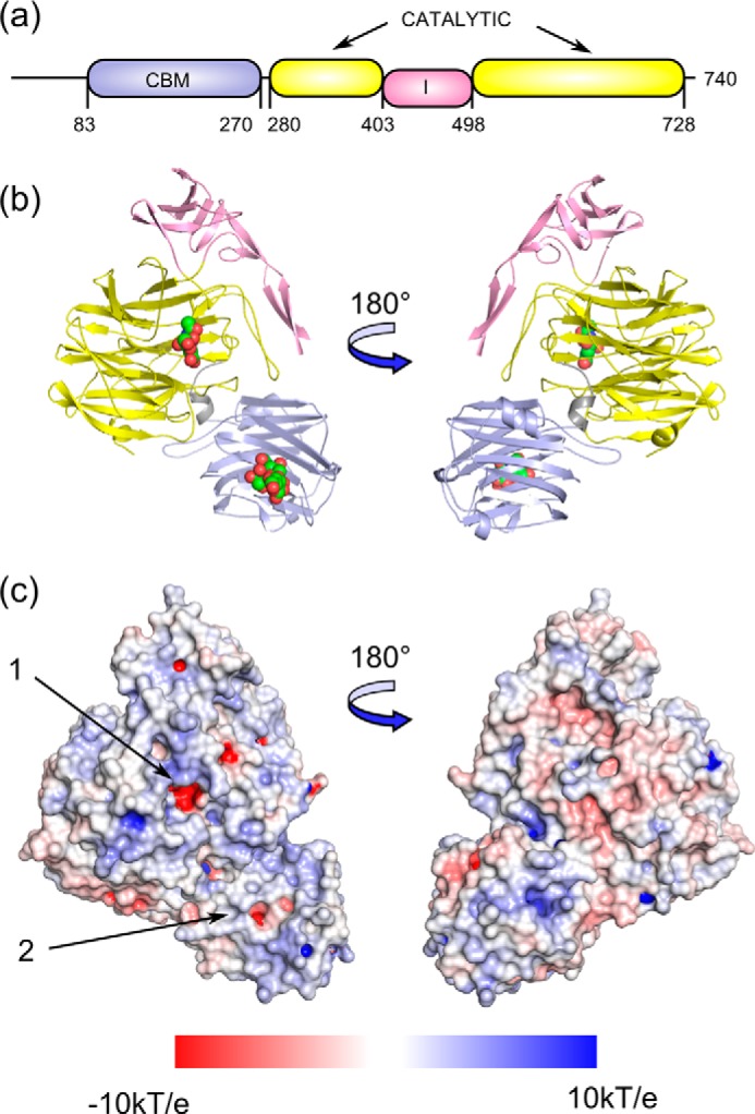
FIGURE 1.

a, NanC domain organization and the amino acid boundaries are shown, with the CBM in light blue, the sialidase catalytic domain in yellow, and the inserted domain in pink. b, in the cartoon representation the bound ligands, Neu5Ac2en in the catalytic domain, and 3′SL in the CBM (PDB code 4YZ5) are shown as spheres. c, an electrostatic surface has been applied to the unbound crystal structure (PDB code 4YZ1) with the catalytic domain active site and the CBM binding site indicated with arrows 1 and 2, respectively. Electrostatic potentials (units of kT/e from −10 to 10) were calculated by APBS (76) and visualized on the solvent-accessible surface by the program PyMOL (77).
