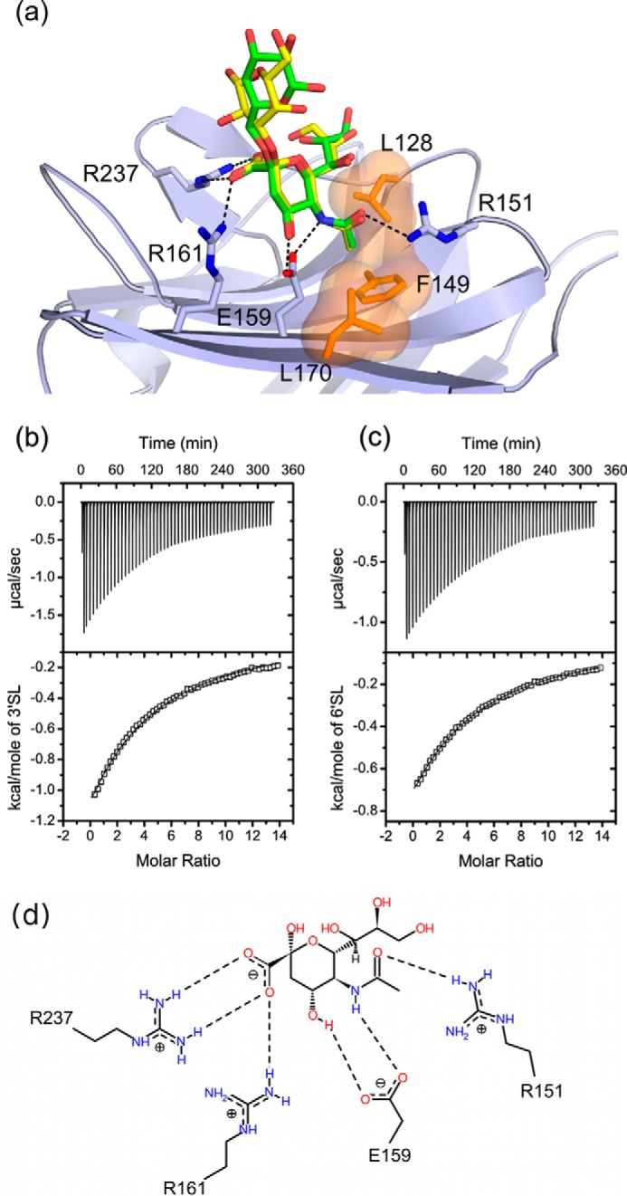
FIGURE 2.

a, the CBM binding site of NanC (light blue) with superimposed bound 3′SL (yellow; PDB code 4YZ5) and 6′SL (green; PDB code 4YW2) molecules shown with selected interacting residues. Dashed black lines indicate hydrogen-bonding interactions and a semitransparent surface (orange) applied to Leu-128, Phe-149, and Leu-170 indicates the hydrophobic pocket. b and c, ITC experiments assessing the binding of 3′SL and 6′SL, respectively, to subcloned NanC CBM. Based on crystallographic evidence, the binding curve was calculated using a fixed substrate to binding site stoichiometry of 1:1. For 3′SL binding constant K = 677 ± 7.44 m−1, ΔH = −11.4 ± 0.078 kcal/mol, TΔS = −7.54 kcal/mol corresponding to a Kd of 1.48 mm. χ2/DoF = 92.49. For 6′SL binding constant K = 626 ± 4.43 m−1, ΔH = 8.09 ± 0.036 kcal/mol, TΔS = −4.26 kcal/mol corresponding to a Kd of 1.60 mm. χ2/DoF = 16.78. d, a schematic representation of the interactions between Neu5Ac and the CBM binding site (PDB code 4YW1) visualized using Poseview (78).
