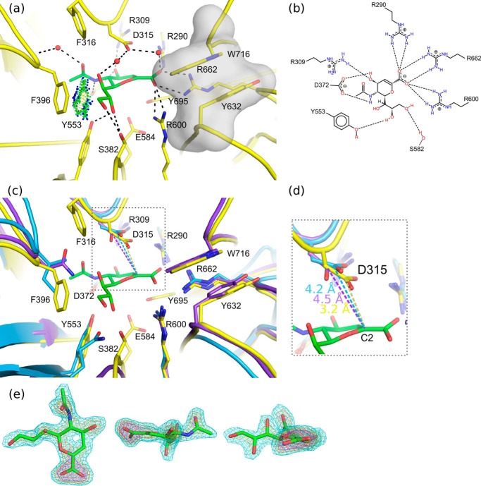
FIGURE 4.
Neu5Ac2en binding. a, the NanC active site (yellow) is shown with Neu5Ac2en (green) bound (PDB code 4YW3). Predicted hydrogen bonding interactions are shown with black dashed lines. A hydrophobic interaction between the C9 and Phe-396 is indicated with green and blue spheres (Van der Waals contacts generated using PROBE (79)). The hydrophobic stack providing specificity for α2,3-linked substrates is highlighted with a semitransparent gray surface. b, a schematic representation of the interactions between Neu5Ac2en and the active site. c, for comparison the active sites of NanA (blue) and NanB (purple) are aligned onto NanC. d, a close-up view of the distances in Å between the Neu5Ac2en anomeric carbon and conserved acid base aspartic acid. e, three orientations of the bound Neu5Ac2en ligand are shown. The Fo − Fc omit electron density map is contoured at sigma levels of 3 (cyan), 5 (orange), and 7 (magenta) carved at a distance of 1.6 Å.

