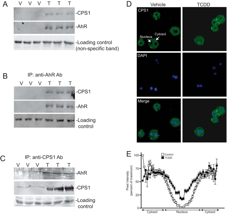
FIGURE 2.
AhR interaction and nuclear translocation of CPS1. A, whole liver nuclear extracts (50 μg) isolated from C57BL/6 mice treated with 20 μg/kg TCDD (T) or vehicle (V) for 2 h were subjected to immunoblotting. The results are for independent animals. B, vehicle- (V) and TCDD-treated (T) mouse liver nuclear protein (1 mg) was immunoprecipitated (IP) with an antibody against AhR and immunoblotted for the AhR and CPS1 as independent animals in triplicate. C, reciprocal immunoprecipitation using a CPS1 antibody followed by immunoblotting as described for B. D, indirect immunofluorescence confocal microscopy on vehicle (DMSO) and TCDD-treated primary hepatocytes stained for CPS1 (green) and DAPI (blue). E, densitometric quantitation across individual hepatocytes was performed to measure nuclear and cytosolic CPS1 staining and plotted as percentages of pixel intensity. The data represent n = 47 cells for vehicle controls and n = 46 for TCDD-treated cultures (in 12 random fields from two independent experiments).

