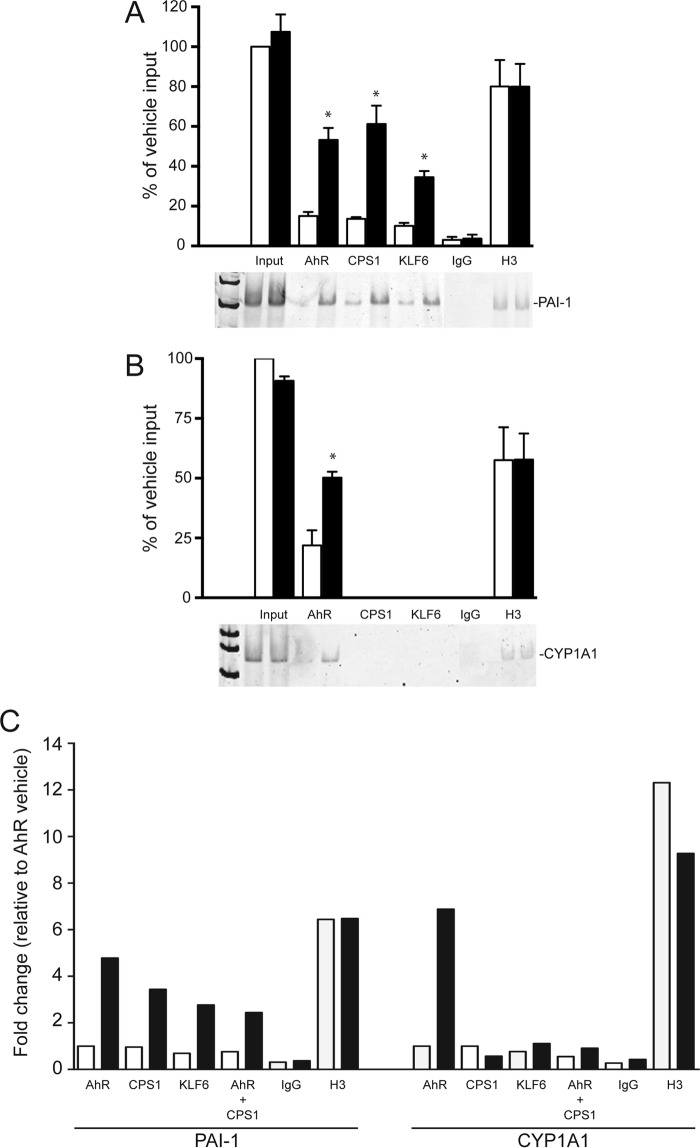
FIGURE 3.
TCDD-dependent AhR and CPS1 binding to the PAI-1 promoter. ChIP assays were performed on liver tissue isolated from vehicle-treated (open bars) and TCDD-treated (solid bars) floxed mice (20 μg/kg for 2 h). Antibodies against AhR, CPS1, KLF6, H3 (positive control), and IgG (negative control) were used to immunoprecipitate target proteins. PCR primers against the PAI-1 and CYP1A1 promoters flanking NC-XRE and XRE sites, respectively, were used to amplify genomic DNA. PCR products were electrophoretically fractionated and stained with SYBR green. A and B, quantitation of PCR products against the PAI-1 promoter (A) and CYP1A1 promoter (B) from vehicle (open bars) and TCDD-treated mice is presented as a percentage of vehicle input DNA (n = 3, average ± S.E.). *, p < 0.05. C, quantitative PCR was performed on DNA isolated from vehicle- and TCDD-treated mouse livers in a sequential re-ChIP experiment using antibodies against the AhR, followed by CPS1.

