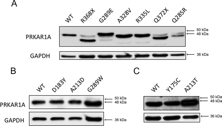
FIGURE 1.
Expression of mutant PRKAR1A subunits analyzed by Western blotting. Cell lysates were prepared from HEK293 cells transfected with plasmids, resulting in the expression of WT PRKAR1A or PRKAR1A carrying the indicated mutations (A, WT, R368X, G289E, A328V, R335L, Q372X, and Q285R; B, WT, D183Y, A213D, and G289W; C, WT, Y175C, and A213T). Soluble proteins were electrophoresed into 10% SDS-polyacrylamide gels and transferred to nitrocellulose membranes, and PRKAR1A (top gels) and GAPDH (bottom gels) were detected by chemiluminescence after sequential treatment of the membranes with specific primary antibodies, peroxidase-conjugated secondary antibodies, and ECL substrate. The arrows show the migration of proteins of the indicated molecular mass (kDa).

