FIGURE 8.

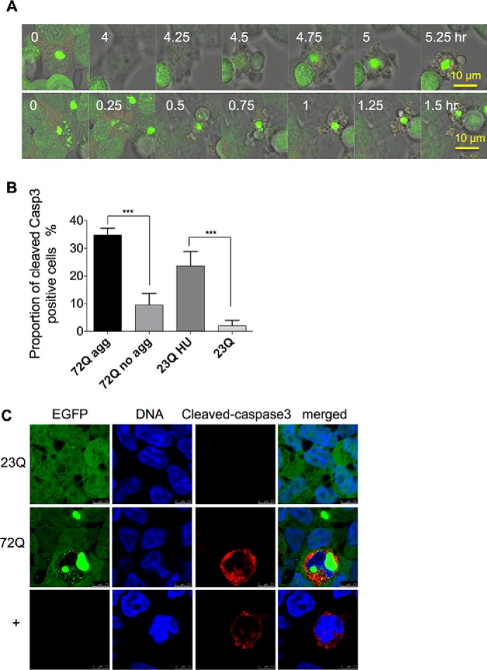
Aggresome-containing cells undergo apoptosis. A, time-lapse confocal microscopy of apoptosis in two representative aggresome-containing cells. The process can take from 2 h to 5 h. Live samples of long term-induced (3 months) EGFP-HDQ72 cells were recorded by confocal microscopy, which demonstrated three apoptotic cells among 12 aggresome-containing cells. B, quantitation of cells positive for cleaved caspase-3. Apoptosis in cells expressing EGFP-HDQ72 over the long term (3 months) is strongly correlated with the presence of aggresomes. EGFP-HDQ23 cells treated with hydroxyurea (1.5 mm for 8 h) act as the positive control, and EGFP-HDQ23 cells act as the negative control. Three independent experiments were carried out, with ∼200 cells assessed per experiment in each sample (EGFP-HDQ72, EGFP-HDQ23, and EGFP-HDQ23 hydroxyurea (HU)). The chart represents the mean ± S.D. Samples were analyzed by binary logistic regression. ***, p < 0.001. C, immunostaining of cleaved caspase-3 in long term-induced (3 months) EGFP-HDQ72 cells showed that a proportion of EGFP-HDQ72 cells undergoes apoptosis. Negative control: long term induced EGFP-HDQ23 cells. Positive control: uninduced cells treated with hydroxyurea (1.5 mm for 8 h).
