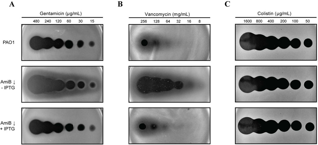
Figure 5. Antibiotic susceptibility of AmiB-depleted cells.
A. Gentamicin spotting assay. Overnight cultures of PAO1 [WT] and BPA46 [ΔamiB (PTOPLAC::amiB)] were diluted 1:100 in LB supplemented with 1mM IPTG and allowed to grow at 30°C for 3h. Following two washes with fresh LB, the cells were resuspended in LB lacking IPTG and allowed to grow for another 2h. They were then normalized to OD600 of 0.4, diluted 1:40 in molten LB 0.8% agar either containing or lacking 1mM IPTG as indicated, and distributed over LB plates. After allowing 30min at room temperature for the agar to solidify, 5µL aliquots of gentamicin solutions at the indicated concentrations were spotted onto the surface of the plates. Plates were incubated at room temperature (~22°C) for 2d and then photographed.
B. Vancomycin spotting assay. Cells were treated the same as in (A), except that 10µL aliquots of vancomycin solutions at indicated concentrations were spotted onto the plates.
C. Colistin spotting assay. Cells were treated the same as in (A), except that 5µL aliquots of colistin solutions at indicated concentrations were spotted onto the plates.

