FIGURE 3.
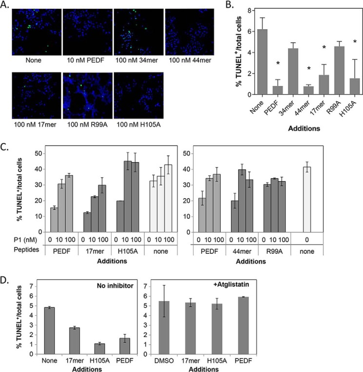
Protective effects of PEDF peptides in cultured cells. R28 cells in serum-free media were treated with PEDF-derived peptides for 48 h. Cytoprotection was assayed by counting TUNEL-positive nuclei (green). A, representative images from each condition show merged TUNEL+ nuclei and Hoechst stained nuclei (blue, total cells). Concentrations of peptides were as indicated. B, quantification of the protective effects by PEDF peptides. Each bar corresponds to the average of the percentage of TUNEL-positive nuclei per total number of cells from triplicate wells ± S.D. (error bars) with treatment as indicated in A. *, p < 0.03. C, blocking of the protective effects of PEDF peptides by P1 peptide. Treatments were with PEDF or peptides at 10 nm mixed with P1 peptide at the indicated concentrations. D, cells were pretreated with atglistatin (3.5 μm) in DMSO or DMSO (vehicle control) for 1 h before a 48-h treatment with or without PEDF peptides and protein at 10 mm (indicated). Cells were fixed and processed for TUNEL staining and counterstained with Hoechst dye for the nucleus. Quantification of TUNEL-positive nuclei is shown. Each bar corresponds to the average of percentage of TUNEL-positive nuclei per total number of cells from duplicate wells per assay ± S.D. Each plot shows assays performed with an individual batch of R28 cells. PEDF and no additions were positive and negative controls, respectively.

