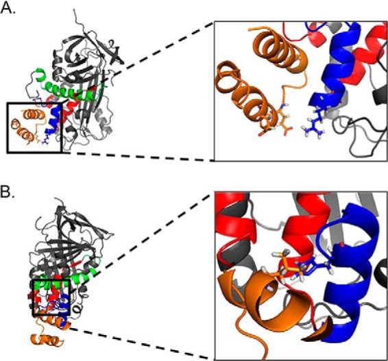
FIGURE 7.

PEDF amino acids that modify binding to the P1 fragment of the PEDF receptor. P1 peptide (orange) interacts with the solvent accessible 17-mer peptide region (blue) within the neuroprotective 44-mer peptide region (red). A, the inset shows the close proximity of the positively charged amino acid Arg99 (blue) of PEDF and two negatively charged amino acids Glu230 and Glu236 (orange) of the LBD of PEDF-R that could be important for binding. B, the molecules are rotated 90° to the right on the y axis and 15° forward on the z axis relative to A. The inset shows the close proximity of the hydrophilic His105 (blue) to the hydrophobic Leu222 (orange) in the P1 fragment. Amino acids Arg99 and His105 of PEDF and Glu230, Glu236, and Leu222 are shown with side chains in sticks.
