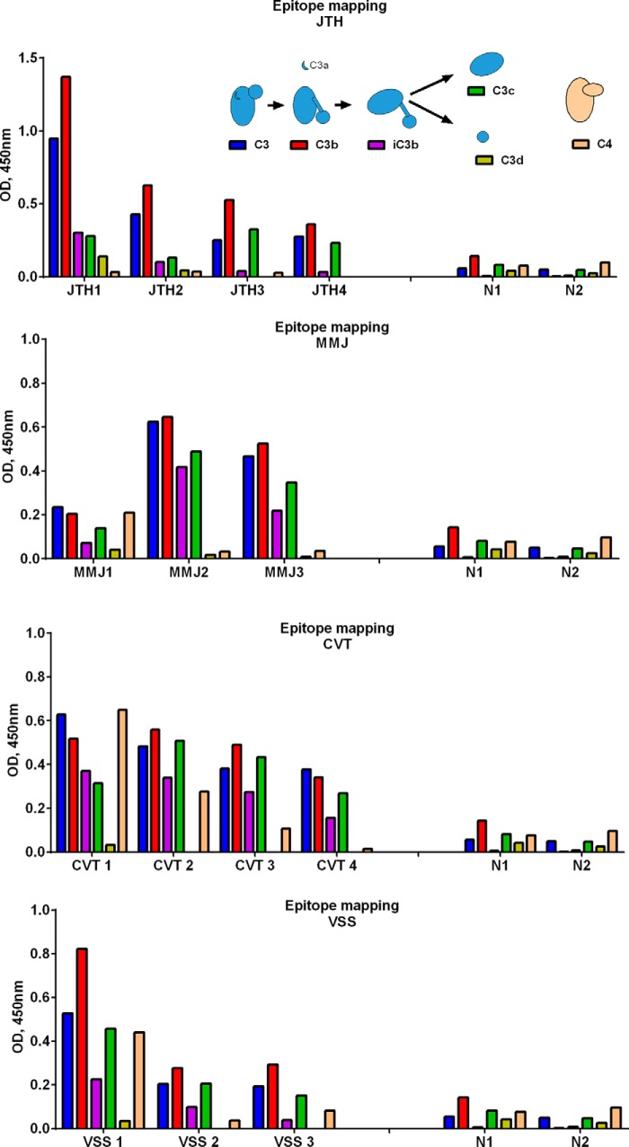
FIGURE 4.
Epitope mapping of the anti-C3 antibodies. The binding of IgG from patients' plasma to C3 and its different fragments (C3b, iC3b, C3c, and C3d) and to C4 was measured by ELISA. The inset gives schematic representation of C3 and the cleavage process to obtain the different functional fragments used for the epitope mapping. Results are presented for the patients JTH (A), MMJ (B), CVT (C), and VSS (D).

