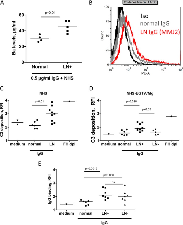
FIGURE 7.
Effect of the anti-C3 autoantibodies on complement activity in normal human serum. A, generation of Ba after incubation of IgG from heathy donors and anti-C3 antibody-positive patients with a normal human serum in the presence of EGTA-Mg. B–D, C3 deposition on the endothelial cells from normal human serum, supplemented with IgG from LN patients or healthy donors (Normal), measured by flow cytometry. B, representative histogram from FACS analysis of the deposition of C3 in the presence of anti-C3 autoantibody-positive IgG. C, C3 deposition from normal serum, supplemented with IgG from all positive patients for whom a sufficient amount of IgG was available (n = 9), compared with IgG from healthy donors (n = 6). D, C3 deposition on endothelial cells from normal human serum, supplemented with EGTA-Mg2+ (to block the classical and lectin pathways) and IgG from healthy donors (n = 6) or LN patients, positive (LN+; n = 9) or negative (LN−; n = 5) for anti-C3 autoantibodies. Factor H-depleted serum served as a positive control, and the absence of IgG (medium only) was the negative control for C3 deposition in C and D. E, the presence of anti-endothelial cell IgG antibodies in patients with LN, positive (LN+) or negative (LN−) for anti-C3 autoantibodies, compared with healthy donors. The statistical analyses were performed by Mann-Whitney t test. HUVEC, human umbilical vein endothelial cells.

