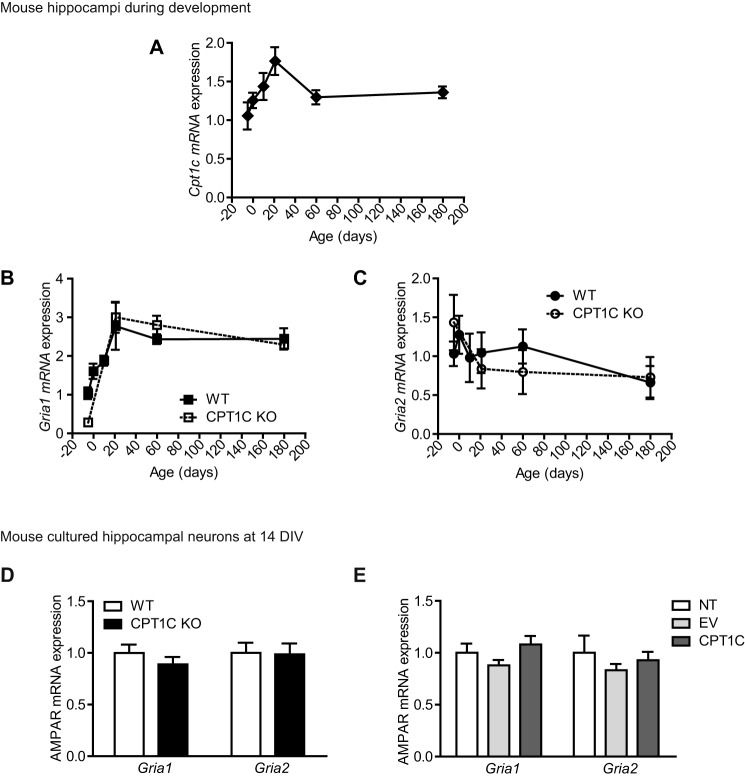
FIGURE 6.
Gria1 and Gria2 mRNA levels in CPT1C KO animals. A–C, hippocampi were dissected from the following WT and CPT1C KO specimens: 17-day post coitus fetuses and 0-, 10-, 21-, 60-, and 120-day-old mice. Cpt1c (A), Gria1 (B), and Gria2 (C) mRNA levels were determined by real-time PCR. Gapdh was used as a housekeeping gene. Graphs show the mean ± S.E. (error bars) of 3 animals/group. D, AMPAR mRNA levels detected at 14 DIV in WT or CPT1C KO hippocampal neurons in culture. Results are shown as mean ± S.E. in three independent experiments performed in duplicate. E, Gria1 and Gria2 levels in WT neurons infected with lentiviral CPT1C. Results are shown as mean ± S.E. in two independent experiments performed in triplicate.

