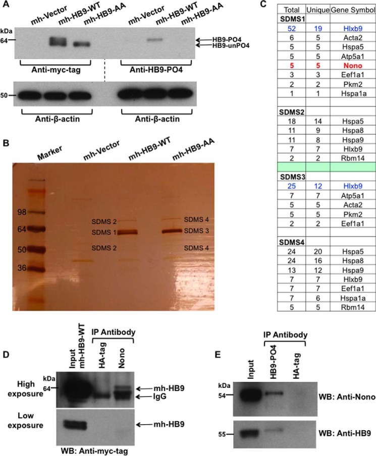
FIGURE 1.
Identification of Nono as a phospho-HLXB9 interacting protein. A, overexpression of HLXB9 shows both its phosphorylated and unphosphorylated isoform. WCE were prepared from MIN6-4N cells transfected with myc-His-tag empty vector (mh-Vector) or plasmids expressing myc-his-tagged HLXB9 (mh-HB9-WT) or the phospho-dead mutant of HLXB9 with alanine substitution at Ser-78 and Ser-80 (mh-HB9-AA). WCE were run on the same gel to generate two Western blots to probe with anti-myc-tag or anti-HB9-PO4 (phospho-HLXB9 antibody). To analyze the bands, the blots were placed side-by-side (indicated by the dotted line). The top band of the doublet in the lane marked mh-HB9-WT corresponds to phospho-HLXB9 because it is not detected with the myc-tag antibody in mh-HB9-AA, and it is detected specifically with anti-HB9-PO4. The bottom band of the doublet corresponds to the unphosphorylated isoform of HLXB9 (HB9-unPO4) because it is not detected with anti-HB9-PO4. β-Actin was used as the loading control. B, large scale co-IP shows the two isoforms of HLXB9 and co-immunoprecipitating proteins. Silver-stained gel of proteins separated on SDS-PAGE after large scale co-IP with a myc-tag antibody using WCE prepared in A. As also seen by Western blot analysis in A, the bands marked SDMS1 and SDMS3 show the doublet in mh-HB9-WT Co-IP (PO4-HB9 and unPO4-HB9) and a single band in mh-HB9-AA corresponding to unPO4-HB9. Bands marked SDMS1, SDMS2, SDMS3, and SDMS4 (that were absent in the mh-Vector lane) were excised from the gel and subjected to mass spectrometry analysis. C, Nono uniquely co-immunoprecipitates with phospho-HLXB9. The number of peptides (total and unique) in the bands excised from the gel shown in B and their corresponding proteins is shown. Several proteins were present in both the mh-HB9-WT and mh-HB9-AA immunoprecipitates. The protein Nono emerged as a phospho-HLXB9-specific partner found only in the co-IP of mh-HB9-WT and not in the co-IP of the phospho-dead mutant of HLXB9 (mh-HB9-AA). D, overexpressed HLXB9 can Co-IP with endogenous Nono. Western blot (WB) probed with anti-myc-tag showing specific co-IP of HLXB9 with Nono using WCE of MIN6-4N cells transfected with mh-HB9-WT. Anti-HA-tag was used as a negative control. Higher exposure (top panel) and lower exposure (bottom panel) of the blot are shown to clearly visualize the input HLXB9 bands. IP, immunoprecipitation. E, endogenous phospho-HLXB9 can co-immunoprecipitate with endogenous Nono. Western blots probed with anti-Nono and anti-HB9 show specific co-IP of endogenous Nono with endogenous phospho-HLXB9 using WCE of MIN6-4N cells. An anti-HA-tag was used as a negative control.

