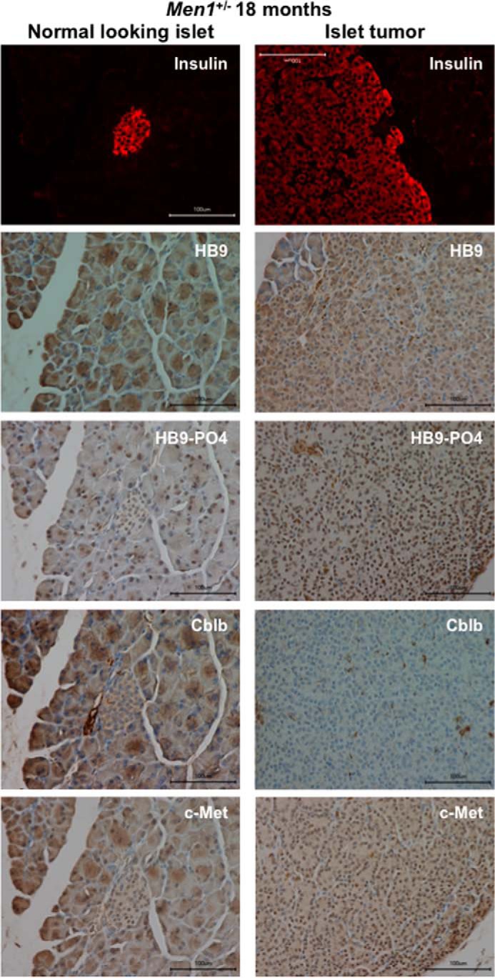
FIGURE 7.

Increased phospho-HLXB9, decreased Cblb, and increased c-Met in insulinoma from the conventional mouse model of menin loss (Men1+/−). Shown are images of immunofluorescence for insulin and IHC for the indicated proteins in the pancreas section of an 18-month-old Men1+/− mouse. Insulin staining shows the location of the normal-looking islet (panels on the left) and the large islet tumor that covers the entire viewing field (panels on the right). Compared with a normal-looking insulin-positive islet in the same section, the insulin-positive islet tumor shows increased nuclear staining for HB9 and HB9-PO4 and increased nuclear and cytoplasmic staining for c-Met but almost no staining for Cblb (cytoplasm).
