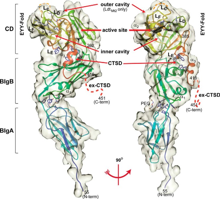
FIGURE 1.
Crystal structure of LdtMt5. Left, apo-LdtMt5; right, rotated 90°. LdtMt5 is composed of two BIg domains and a CD. The semitransparent volume is surface-accessible by a 3.5-Å radius probe. Residues of the active site (His342, Thr357, Asn358, and Cys360) are represented as sticks within the CD. Tryptophan residues of the CTSD are also represented as sticks and interact with a hydrophobic patch at the interface of the BIgB domain. The prominent outer cavity that is observed in LdtMt2 is absent in apo-LdtMt5 but is indicated as a reference (10). The LdtMt5 secondary structure schematic is colored as a rainbow from blue (N terminus) to red (C-terminus). Orange dashes represent the disordered portion of loop LC, and red dashes represent the disordered ex-CTSD. This figure was made using Chimera (19).

