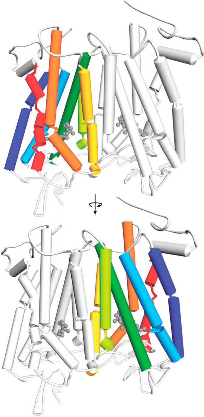
FIGURE 1.

Model of the Rho dimer. Transmembrane helices in each Rho monomer are colored as follows: TM1, dark blue; TM2, light blue; TM3, dark green; TM4, light green; TM5, yellow; TM6, orange; and TM7, red. The same color code is also used in Figs. 2–5 and 7–9. The dimer-contacting surface is based on modeling studies involving helices TM4 and TM5 (28). The second monomer within the Rho dimer is colored light gray.
