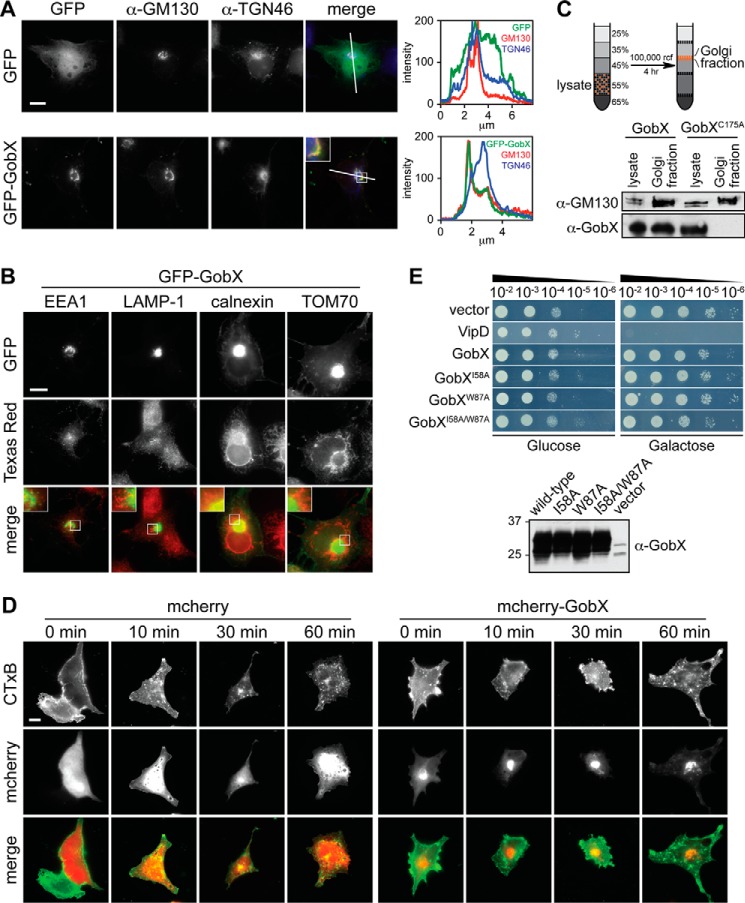
FIGURE 3.
GobX localizes to the Golgi region. A, intracellular localization of GobX. Transiently transfected COS-1 cells producing either GFP (control) or GFP-GobX were labeled with antibody directed against the medial Golgi marker GM130 or the trans-Golgi network marker TGN46 and imaged by fluorescence microscopy. Scale bar, 2 μm. The pixel intensity of the green (GFP), red (GM130), and blue (TGN46) fluorescent signals along the line indicated in the image is shown at the right. Enlarged view of the boxed region is shown in the inset. B, GobX does not co-localize with endosomes, lysosomes, ER, or mitochondria. COS-1 cells producing GFP-GobX were labeled with antibody directed against different organelle markers and imaged by fluorescence microscopy. EEA-1, early endosome; LAMP-1, lysosome; calnexin, ER; TOM70, mitochondria. Scale bar, 2 μm. The boxed regions are magnified in insets for each panel. C, GobX was enriched in the Golgi fraction in infected macrophages. RAW264.7 macrophages challenged with Lp02 producing HA-GobX or HA-GobXC175A was fractionated by sucrose discontinuous gradient centrifugation. The total cell lysate and the fraction enriched in Golgi membranes (45/35% interface) were probed by immunoblot using anti-GM130 and anti-GobX antibody. D, GobX does not interfere with retrograde trafficking. Transiently transfected COS-1 cells producing either mCherry or mCherry-GobX were incubated with CTxB for 10 min, and CTxB traffic was monitored after 0, 10, 30, and 60 min within the cell. Scale bar, 2 μm. E, GobX does not interfere with yeast growth. Serial dilutions (10−2–10−6) of S. cerevisiae INVSc1 cells containing plasmids encoding GobX or GobX variants with attenuated E3 ligase activity were plated on repressing (glucose) or inducing (galactose) synthetic medium plates and analyzed for growth after 48 h. VipD served as a growth defect control (20). Production of GobX and GobX variants in INVSc1 cells was analyzed in liquid culture of synthetic media containing 2% (w/v) galactose and detected on an immunoblot using anti-GobX antibody. All immunofluorescence images, immunoblots, and yeast growth assays are representative of at least two independent experiments with similar outcomes.

