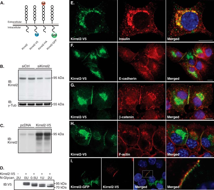
FIGURE 1.
Kirrel2 is predominantly expressed at the cell junctions and co-localizes with adherens junction proteins. A, schematic representation of wild type N- and C-terminally tagged Kirrel2 proteins. B, Western blot of MIN6 cells transfected with control or Kirrel2 targeting siRNAs using an antibody against the N-terminal domain of Kirrel2. γ-Tub, γ-tubulin. C, immunoblots (IB) of MIN6 cells transfected with control (pcDNA) or Kirrel2-V5 expression vectors using anti-Kirrel2 antibody. D, lysates of MIN6 cells transfected with control (−) or Kirel2-V5-expressing (+) plasmids were treated with indicated units of N-glycanase enzyme and immunoblotted with anti-V5 antibodies. E–H, MIN6 cells expressing Kirrel2-V5 were immunolabeled with indicated antibodies or phalloidin (F-actin). I, MIN6 cells were transfected with Kirrel2-V5 or Kirrel2-GFP vectors. After 16 h, the two cell populations were trypsinized, mixed, and cultured on chambered microscopy slides for 16 h prior to immunolabeling with anti-V5 antibodies. Images were obtained by confocal microscopy. Bar, 10 μm.

