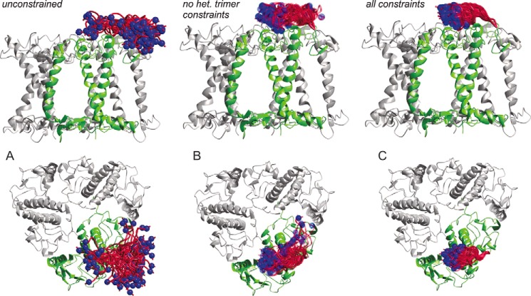
FIGURE 10.
Model of 25 possible conformations of amino acid residues 3–13 of a LHCII monomer in a trimeric assembly viewed from the membrane plane (top) and from above the membrane normal (bottom). Blue spheres show a space-filling model of the Cα atom of residue 3. A, model without any restraints. B, restraints based on data published in Ref. 6. C, restraints based on data from Ref. 6 and heterogeneous trimer measurements.

