
FIGURE 1.
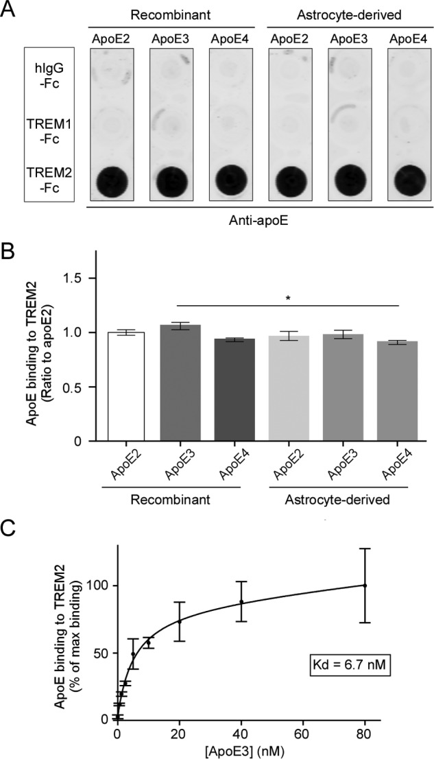
ApoE specifically binds to TREM2 with high affinity. A, representative dot blot of apoE binding to TREM2. A nitrocellulose membrane was spotted with 200 ng each of recombinant human IgG Fc region (hIgG-Fc), recombinant human TREM1, or TREM2 extracellular region conjugated to Fc (TREM1-Fc and TREM2-Fc). Membrane strips were then incubated with 10 nm recombinant or immortalized astrocyte-derived apoE2, -E3, or -E4. Bound apoE was detected using biotinylated anti-apoE antibody. B, quantification of three independent dot blots. Data are plotted as mean ± S.E. (n = 3, one-way analysis of variance with Tukey post hoc analysis, *, p < 0.05). C, saturation binding curve and dissociation constant (Kd) of apoE3 binding to TREM2-Fc. Membrane strips spotted with hIgG-Fc, TREM1-Fc, or TREM2-Fc were incubated with increasing concentrations of recombinant apoE3, and bound apoE was detected using appropriate antibodies. The curve fit and Kd were derived using GraphPad Prism nonlinear fit for one site total binding. Data are plotted as mean ± S.E. (n = 3).