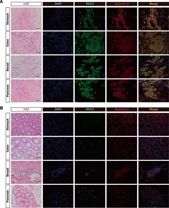
FIGURE 4.
Distributions of MUC1 and galectin-3 in various tumor and nonmalignant tissues. A and B, sections of paraffin-embedded human tumor (A) and nonmalignant (B) tissues (stomach, colon, breast, and pancreas) were stained with hematoxylin and eosin (H&E), DAPI (blue), and the same combinations of antibodies as described in Fig. 3 (MUC1-ND, green; galectin-3, red). Image magnification is ×200. Scale bars, 100 μm.

