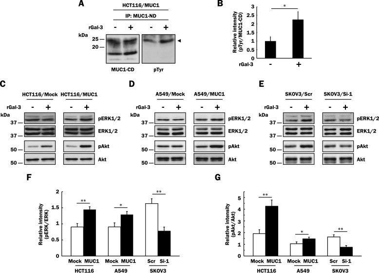
FIGURE 5.
Enhanced phosphorylation of ERK1/2 and Akt through the binding of galectin-3 to MUC1. A, after galectin-3 retained on the cell surface had been excluded as described under “Experimental Procedures,” HCT116/MUC1 cells were treated with recombinant galectin-3 (40 μg/ml, rGal-3 +) or PBS (vehicle, rGal-3 −) for 4 min, and then MUC1 was immunoprecipitated (IP) from lysates of HCT116/MUC1 cells with anti-MUC1-ND antibodies. The immunoprecipitates were subjected to SDS-PAGE, followed by Western blotting and detection with biotin-conjugated anti-phosphotyrosine (pTyr) and anti-MUC1-CD antibodies. The arrowhead in the right panel indicates tyrosine-phosphorylated MUC1-CD. B, the intensities of the bands in Fig. 5A were determined with ImageJ. The ratio of tyrosine-phosphorylated MUC1-CD (pTyr) to MUC1-CD in galectin-3-treated HCT116/MUC1 cells was determined when that in non-galectin-3-treated HCT116/MUC1 cells was taken as 1 (means ± S.D., n = 3). *, p < 0.05. C–E, after exclusion of galectin-3 from the cell surface as described above, HCT116/Mock, HCT116/MUC1 (C), A549/Mock, A549/MUC1 (D), SKOV3/Scr and SKOV3/Si-1 (E) cells were treated with recombinant galectin-3 (40 μg/ml, rGal-3 +) or PBS (vehicle, rGal-3 −) for 10 min, and then the lysates were subjected to SDS-PAGE, followed by Western blotting and detection with anti-phospho-ERK1/2 (pERK1/2), anti-ERK1/2, anti-phospho-Akt (pAkt), and anti-Akt antibodies. F and G, the intensities of the bands in Fig. 5 (C–E) were determined with ImageJ. Each histogram shows the ratios of pERK1/2 to ERK1/2 (F) and pAkt to Akt (G) in each galectin-3-treated cell type when the ratios obtained from each non-galectin-3-treated cell type were taken as 1 (means ± S.D., n = 3). *, p < 0.05; **, p < 0.01.

