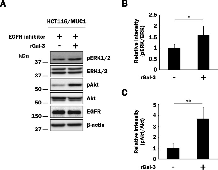
FIGURE 6.
Influence of an EGFR inhibitor on phosphorylation of ERK1/2 and Akt through the binding of galectin-3 to MUC1. A, after exclusion of galectin-3 from the cell surface as described in Fig. 5A, HCT116/MUC1 cells were preincubated with an EGFR inhibitor (10 μm) for 1 h and subsequently treated with recombinant galectin-3 (40 μg/ml, rGal-3 +) or PBS (vehicle, rGal-3 −) for 10 min. Phosphorylated ERK1/2, total ERK1/2, phosphorylated Akt, and total Akt were determined as described in Fig. 5 (C–E). Concomitantly, EGFR and β-actin were also determined. B and C, the intensities of the bands in Fig. 6A were determined as described in Fig. 5 (F and G). The ratios obtained from non-galectin-3-treated HCT116/MUC1 cells were taken as 1 (means ± S.D., n = 4). *, p < 0.05; **, p < 0.01.

